The Department of Gynecological Oncology
Tumors occupy a significant place among female genital diseases. They appear as a result of the pathological transformation of tissue cells that leads to loss of their proper function, unrestricted growth and uncontrolled multiplication. Neoplasms of female genitals include transformation of ovarian, endometrial, cervical, vaginal and vulvic tissues, as well as tumors of the placental tissue during pregnancies. Tumors of the female genital tract may present a great danger to female patients, and therefore, they require early diagnosis, immediate attention and urgent treatment.
The Department of Gynecological Oncology of the medical center at Tel-Ha Shomer Hospital is an affiliate of Sackler Tel-Aviv University Medical School. The Department is at the forefront of the gynecological oncology field regularly introducing new methods to diagnose and treat female genital tumor formations. It is also constantly recognized as a leader in academic support, medical research studies, and its participation in international conferences.
The patients' need for an integrated approach of treating tumors has led to the creation of the Department of Gynecological Oncology, which brings together professionals from different specialties, including gynecologists, oncologists, oncology nurses and social workers, into one multidisciplinary team. The Department treats complications caused by gynecological neoplastic diseases, minimizes the side effects of treatments, and monitors infertility treatments and problem pregnancies.
Research conducted at the Department
Treatment of gynecologic cancer involves combined knowledge of gynecology and oncology, with specialty in treating gynecological disorders and infertility. For this reason, doctors and scientists of the Department study cancer biology of the female reproductive system and the clinical courses of diseases. The scientific team is fully equipped to study all methods of treatment traditionally used in such cases, and is constantly increasing their knowledge in the fields of surgery, chemotherapy and radiation, as well as innovations in medical research.
Key research interests of the medical research staff include evaluating the treatment of uterine, cervical and vaginal tumors, clinical characteristics of tumors at their transition from benign to malignant tumors. Young women admitted with a diagnosis of a malignant uterus tumor receiving a traditional treatment get always checked the effectiveness and efficiency of their therapy. The Department also conducts advanced genetic research, with the assessment of the role of mutations in BRCA 1-2 and the RAS oncogenes in gynecologic neoplasms. These studies are conducted in collaboration with the Oncological Genetics Unit led by Professor Eitan Friedman, and with Professor Yoel Kellogg from the Neurobiochemistry Department of Tel Aviv University.
The medical staff of Gynecology Department includes:
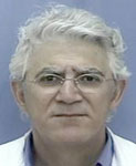
Professor Gilad Ben Baruch - Head of the Department, Acting Head of the Joseph Buchman Center for Obstetrics and Gynecology with a specialty in general gynecology and gynecological oncology |
 |
Dr. Jacob Korah - a Senior Doctor with specialty in gynecology, obstetrics, gynecology oncology |
 |
Dr. Mario Bayner - a Senior Doctor with specialty in gynecology, obstetrics, gynecology oncology, laparoscopic gynecology, and colposcopy of the cervix |
 |
Dr. Nissim Zmira - a Senior Doctor with specialty in gynecology and obstetrics |
 |
Sharon Praster - head nurse |
 |
The entire medical staff has received special academic training in the field of gynecological oncology. Social workers and professional staff are involved to support patients suffering from cancer to ensure that the patients' wellbeing is supported in every way.
Diagnosis
The main scientific and medical activities at the Department of Gynecological Oncology include the state-of-the-art and highly informative diagnostic methods. Neoplastic markers are used to detect malignant tumors of the ovaries and the uterus. Depending on the results of primary observations made by a specialist, and after verifying the initial diagnosis with tumor markers, further examination procedures such as the following are often recommended and performed:
- colposcopy
- hysteroscopy
- X-ray
- virtual computer modeling biopsy
- laparoscopy
- isotope tests
- computed tomography (CT)
- ultrasound tests involving complex magnetic resonance techniques, such as positron emission tomography (PET). PET is an advanced technology to find a tumor, if there is strong suspicion.
PET Scans
PET scans involve injecting of radioactive labeled molecules of natural compounds, such as glucose, into the body to produce an image of internal structures that physicians can use for better diagnosis. The injected fluid spreads throughout the body and emits positively charged particles that create an image on film. Positron emission tomography works because cancerous cells absorb a greater amount of glucose in comparison to healthy cells. Therefore, cancerous cells accumulate more of the radioactive glucose than non-transformed cells do. Because of this, the tumor in the picture stands out from the healthy tissue in color and brightness.
Compared to computer and magnetic resonance imaging (MRI), which gives an idea of the form and structure of organs and tissues, PET shows how they function, and thus reveal the pathology at an earlier stage. Furthermore, PET allows the doctor to view the whole body together with other organs or specific areas being scanned.
PET is a very informative and state-of-the-art research and diagnostic techniques. By cancer patients, it enables an early detection of disease, the identification of metastases, the evaluation of the chemotherapy effectiveness, and the knowledge of when to change the treatment regimen.
No other diagnostic techniques, including ultrasound, X-ray, and nuclear medicine, allows such a precise and rapid diagnosis. A physician may also order a CT scan along with PET to get a clearer and more complete picture of the diseased areas.
Treatments
Diagnosis and evaluation tests are necessary to assess the patient''s condition and to identify the most effective treatment strategy of cancer at the very early stages of disease or of metastases spread. Such treatments include:
- Surgery removing a part of or the entire affected organ. The operation may use conventional or laparoscopic techniques, depending upon the opinion of the surgeon.
- Chemotherapy using modern drugs with the mildest side effects, as well as dealing with the side effects themselves.
- Radiotherapy.
After the treatment, the patient is offered a personalized rehabilitation program.
Treatment of Fibroids
The Department provides several methods to treat myomas or fibroids. Myomas are benign uterine tumors that are quite common for women of the childbearing age. In most cases, fibroids are small and cause no trouble, but sometimes they can grow to the size that leads to clinical symptoms like pain, bleeding, or problems during urination. There are different types of fibroid formations. Fibroids can lead to complications during pregnancy and to infertility.
Myoma therapies include:
- prevention of bleeding by drug therapy
- surgical removal of the uterus or fibroids via abdominal surgery
- the removal of fibroids by hysteroscopy (inserting optical fibers) in those rare cases when fibroids are located on the inner surface of the uterus
- an embolization procedure, in which tiny particles are introduced into the uterine arteries to disrupt blood flow to fibroids, and thus, to stop their development
The Department of Gynecological Oncology extensively applies the new, progressive and most efficient method of focused ultrasound (FUS), a technique used to treat myomas. FUS is a noninvasive method that focuses treatment of the tumor by ultrasound. This progressive method makes it possible to avoid hysterectomy or myomectomy (removal of the uterus or a part of the uterus). This method of treatment uses focused ultrasound of high intensity, aimed at raising the temperature inside the fibroids themselves without damaging the surrounding tissues and organs. In order to determine where to focus the beam of ultrasound, magnetic resonance imaging (MRI) scans are used. The FUS procedure consists of three steps: MRI scan before treatment, direct monitoring of the treatment with MRI, and observation after treatment.
The MRI technique uses a magnetic field generated by large and powerful magnets to create anatomical images, and does not involve harmful radiation. The MRI image lets the surgeon see the uterine fibroids and utilize the ultrasound device to raise the temperature precisely in the selected area to a level sufficient to destroy the fibroid tissue. Studies of the ultrasound device have demonstrated its ability to aim the beam of ultrasound directly at the tumor through other tissues. MRI helps to ensure the effectiveness of ultrasound treatments. When properly executed, the tissue around the uterus will not be damaged.
Examination and treatment using FUS.
- A few days prior to treatment, the patient needs to undertake a series of tests starting with a visit to the gynecologist, who will perform sonography and confirm the presence of any fibroid growths. Standard blood tests for pregnancy and Pap smear will be performed, together with the ultrasound scanning and/or MRI, involving intravenous injection of a contrast agent (gadolinium) to show the tumor. The MRI scan can last 45 minutes.
- In the course of FUS treatment, the patient needs to lie still in the setup for MRI for approximately three hours. A catheter may be used for urination. The patient receives a sedative by an intravenous injection to prevent pain and anxiety. This will facilitate the process of focusing the ultrasound beam used for the MRI scan. During the entire treatment process, the patient will be awake.
- The main disadvantages associated with this procedure include pain in the area of treatment and numbness or pain in the back, legs or neck caused by about three hours of lying face down on the stomach. Sedatives will help to ease the uncomfortable conditions. Patients should inform the staff if they are at risk of blood clotting, or if their family history suggests such a risk. To prevent thrombosis during treatment, we will provide socks of elastic material.
Advantages of FUS:
- Ability to avoid damage to the uterus as a result of surgical intervention
- Ability to avoid scarring as a result of an invasive surgical procedure
- Low level of risk and complications
- Extremely short period of rehabilitation
- Minimal number of side effects
- Performed in a hospital in the MRI office without any need for hospitalization
- Significant improvement of quality of life
Possible disadvantages of FUS:
- FUS treatment is suitable for most women, but not all. Modern diagnostic techniques allow us to make the correct choice of treatment for the patient. Reducing the size of fibroids occurs gradually, not instantly. Feeling better, usually comes quickly, but again, gradually. Sometimes when uterine myomas are large, there is a need for more than one procedure to achieve the desired result. Because the uterus is not removed, new fibroids may develop, which would require further treatment. Focused ultrasound treatment is not suitable for women with very large uterine masses or multiple fibroids located in remote places.
Eligibility for treatment. The FUS treatment is suitable only for women diagnosed with uterine fibroids. Treatment is not intended for pregnant women and women who:
- suffer from certain chronic diseases and heart disease
- receive treatment for blood thinners
- are sensitive to contrast agents
- cannot undergo MRI scans
- have fibroids too large or inaccessible for treatment
Currently, the FUS method of treatment has been used by more than 6000 women from around the world (Israel, Germany, USA, England and Canada). During the observation period following the treatment (up to six months), there were no complications related to FUS treatment. Women who have received this treatment in recent years have noticed a dramatic relief of symptoms (more than 80% of women, three months after the treatment).


